TREATMENTS

Osteochondral Lesion Repair

Osteochondral Lesion Repair Surgery Sydney

Osteochondral lesion repair is surgery to fix damage to both the smooth joint surface (cartilage) and the underlying bone in your knee, often caused by trauma or conditions like Osteochondritis Dissecans (OCD).

Goal

The surgery aims to restore your knee's smooth surface, relieve pain and catching sensations, and help prevent or delay arthritis.

Techniques

Dr Allom uses techniques like microfracture (stimulating healing) or OATS/mosaicplasty (transferring healthy cartilage and bone) depending on the damage.

Recovery

Rehabilitation takes time, often 9-18 months, involving physiotherapy and gradually returning to activities t o ensure the best outcome.

What is an Osteochondral Lesion?

An osteochondral lesion is a specific type of knee injury affecting two important layers:

Articular Cartilage

This is the smooth, white tissue covering the ends of your bones inside the joint. Think of it as the knee's cushion and gliding surface. It allows for frictionless movement and absorbs shock but has very limited ability to heal itself because it lacks a direct blood supply.

Subchondral Bone

This is the layer of bone directly beneath the cartilage. It provides structural support and, crucially, has the blood supply needed for healing.
When both these layers are damaged in a specific area, it is called an osteochondral lesion or defect. If left untreated, these defects can cause ongoing pain, swelling, mechanical symptoms like catching or locking, and lead to progressive joint deterioration, much like a pothole developing on a smooth road.

Types of Osteochondral Lesions

There are two main types:

Osteochondritis Dissecans (OCD)

In this condition, typically seen in adolescents and young adults, a segment of bone and the cartilage above it loses its blood supply. This segment might become unstable or even break loose completely. The severity depends on whether the fragment is stable (still in place) or unstable/displaced (loose).

Post-Traumatic Osteochondral Defects

These result directly from an injury. A sharp impact, a twisting force during sport (like an ACL tear or kneecap dislocation), or repetitive stress can cause a piece of cartilage and bone to shear off or get damaged.

Common Locations in the Knee

These lesions often occur in specific areas:

Medial Femoral Condyle

The inner side of the thighbone's end. This is the most common site, especially for OCD, and is critical for weight-bearing.

Lateral Femoral Condyle

The outer side of the thighbone's end, often injured during ACL tears.

Patellofemoral Joint

The joint between the kneecap (patella) and the thighbone, frequently damaged during kneecap dislocations.

Tibial Plateau

The top surface of the shinbone. Less common but can occur, sometimes alongside meniscus tears.
Understanding exactly where and how deep the damage is helps determine the best treatment approach.

How Dr Allom Delivers Osteochondral Repair

Dr Allom uses a patient-specific approach, selecting the best surgical technique based on the lesion's size, location, your age, activity goals, and any associated injuries. The aim is always to restore the joint surface as effectively as possible.

Discovery and Diagnosis Phase

Detailed Assessment

Your journey starts with a thorough discussion about your symptoms, injury history, and activity goals. Dr Allom performs a detailed clinical examination to assess pain, swelling, range of motion, and stability.

Advanced Imaging

An MRI scan is usually essential. It provides a detailed picture of the cartilage and bone, showing the lesion's size, depth, stability, and any other injuries like ligament or meniscus tears. X-rays help assess overall joint alignment and bone structure.

Arthroscopic Confirmation

Sometimes, the final assessment happens during surgery using arthroscopy (keyhole camera). This allows direct visualisation and probing of the lesion to confirm stability and guide the precise repair technique.

Planning and Technique Selection Phase

Based on the assessment, Dr Allom discusses the most suitable surgical options with you. Key techniques include:

Microfracture

For smaller defects (<2-4cm²), small holes are made in the underlying bone to release stem cells and stimulate the growth of repair cartilage (fibrocartilage). This is often a first-line treatment.

Osteochondral Autograft Transfer (OATS/Mosaicplasty):

For small to medium defects (1-4cm²) in critical weight-bearing areas, especially in younger, active patients. Healthy cartilage and bone plugs are taken from a non-critical area of your knee and transferred into the defect, like fitting mosaic tiles. This restores the defect with your own hyaline cartilage.

Fragment Fixation (for OCD)

If an OCD fragment is unstable but intact, especially in adolescents, it can often be fixed back into place using special pins or screws, potentially preserving the original cartilage.

(Other options like Allograft)

For very large defects (>4cm²) or revisions, using donor cartilage and bone (allograft) might be considered.
Biological augmentation (like PRP) might be used alongside these techniques to potentially enhance healing.

Implementation and Delivery Phase

Surgical Precision

The chosen technique is performed meticulously, often using arthroscopic (keyhole) methods for microfracture or fragment fixation, or a small open incision for OATS. Precision is key for integrating grafts or stimulating effective healing.

Concurrent Injury Management

If other injuries like ACL tears or meniscus tears are present, these are often addressed during the same operation.

Results and Rehabilitation Phase

Structured Rehabilitation

This is critical. You will work closely with a physiotherapist following a specific protocol based on your surgery. This involves stages of protection (often limited weight-bearing initially), progressive loading, functional training, and eventually, return to activity. Adherence is vital for success.

Monitoring

Follow-up appointments with Dr Allom include progress checks and sometimes imaging (X-ray or MRI) to monitor healing, especially after OCD fixation.

Return to Activity

Timelines vary significantly. Microfracture typically requires 9-18 months for return to sport, while OATS might be slightly faster at 6-12 months. Realistic expectations are important.

Why Choose Dr Allom for Osteochondral Repair?

Choosing the right surgeon is crucial for managing complex knee injuries like osteochondral lesions. Dr Allom offers:
Fellowship-Trained Knee Subspecialist Expertise
With dual international fellowships from the Royal College of Surgeons of England (FRCS) and the Royal Australasian College of Surgeons (FRACS Orth), Dr Allom has dedicated, advanced training specifically focused on knee surgery, including complex cartilage restoration. This depth of expertise goes beyond general orthopaedic training.
Comprehensive Treatment Options
Dr Allom performs the full spectrum of modern cartilage repair techniques, including microfracture, OATS/mosaicplasty, and OCD fragment fixation. He selects the most evidence-based option tailored precisely to your lesion and goals.
Evidence-Based, Personalised Approach
Treatment decisions are grounded in the latest research and your specific circumstances (lesion size, location, age, activity demands). You receive a clear explanation of the rationale behind the recommended approach.
Advanced Arthroscopic Skills
Many procedures can be performed using minimally invasive arthroscopic techniques, potentially leading to faster recovery times compared to traditional open surgery.
Structured Rehabilitation Partnership
Dr Allom collaborates closely with expert physiotherapists, providing clear, technique-specific rehabilitation protocols and objective milestones to guide your recovery safely and effectively.
Regional Accessibility
Offering subspecialist knee expertise across convenient locations in South West Sydney (Gledswood Hills, Campbelltown, Liverpool) and the Mid North Coast (Taree, Forster), saving you travel to Sydney CBD.
Patient-Centred Communication
Dr Allom prioritises clear explanations, shared decision-making, and realistic expectations, ensuring you are fully informed and comfortable throughout your treatment journey.

Frequently Asked Questions

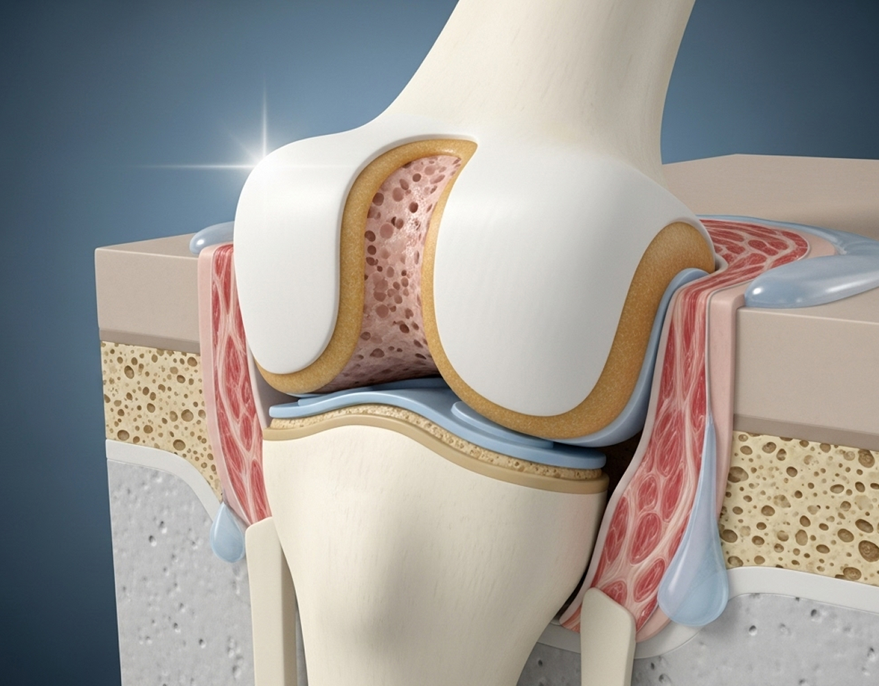
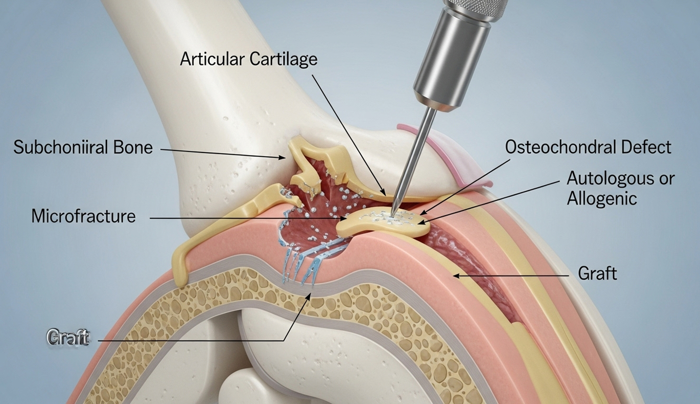
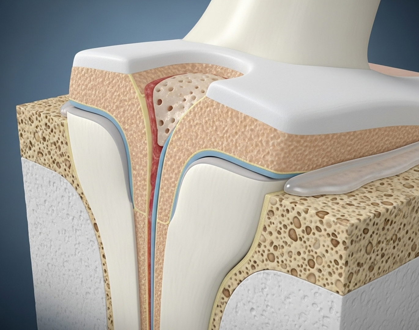
Pure cartilage damage affects only the smooth surface layer (a chondral lesion). An osteochondral lesion involves damage to both the cartilage and the underlying bone. Because the bone has a blood supply, osteochondral lesions sometimes have better potential for healing stimulation (like microfracture) compared to isolated cartilage damage.

Articular cartilage has very poor self-healing ability due to its lack of blood supply. Small, stable defects might not worsen, but they rarely fill in with new, healthy cartilage. Surgery aims to either stimulate a repair response or replace the damaged tissue.

Surgery is often recommended if you have persistent pain or swelling despite non-surgical treatment for 3-6 months, mechanical symptoms like catching or locking, large or unstable lesions seen on MRI, or if your activity goals are limited by the lesion. Small, stable lesions, especially in growing adolescents, might be managed non-surgically first with close monitoring.

There isn't one single "best" treatment. The ideal technique depends heavily on the lesion's size, depth, location, and stability, as well as your age and activity level. Microfracture is common for smaller defects, OATS for medium defects in active people, and fragment fixation for suitable OCD lesions. Dr Allom will discuss the most appropriate evidence-based option for you.

Return-to-sport timelines vary widely depending on the procedure performed and the demands of your sport:

Microfracture

Typically 9-12 months for recreational sports, potentially 12-18+ months for competitive or high-impact sports.

OATS/Mosaicplasty

Generally faster, around 6-9 months for recreational sports, 9-12+ months for competitive levels.

OCD Fixation

Often requires a longer period of protection, with sport return typically around 12-18 months after healing is confirmed.

An osteochondral lesion does increase the long-term risk of developing arthritis in that joint. Surgery aims to restore the joint surface, reduce symptoms, and potentially delay the onset or slow the progression of arthritis compared to leaving the defect untreated. It doesn't eliminate the risk entirely, but it often provides many years of improved function.

Get Started with Osteochondral Repair

If you have ongoing knee pain, catching, or swelling, particularly after an injury or if diagnosed with OCD, an expert assessment is the first step. Dr Richard Allom can provide a precise diagnosis and discuss the most effective treatment options tailored to your knee condition and lifestyle goals.

What to Bring: Please bring your referral letter, all relevant imaging studies (X-rays, CT scans, MRI if performed), and your list of current medications to your consultation.

Convenient Locations in Two Regions

I consult and operate from five locations, providing local access to subspecialist expertise without the need for long-distance travel.

South West Sydney Locations

Phone Number

04 3818 3832

Gledswood Hills

The George Centre, Suite 12, 1A The Hermitage Way

Campbelltown

Centric Park, Level 3, 4 Hyde Parade

Liverpool

Sydney South West Private Hospital, Suite 3.02, 24-40 Bigge Street

Mid North Coast Locations

Phone Number

02 6551 0722

Taree

Mayo Private Hospital Specialist Suites, 2 Potoroo Drive

Forster

Dolphin Suites Consulting Rooms, 33 Breckenridge Street

Book Your Consultation

©2026 Dr Richard Allom. All rights reserved.

Oxford Knee Score Questionnaire

Please answer the following 12 questions about your knee, considering your symptoms over the past 4 weeks. Select one answer for each question.