CONDITIONS

Osteochondral Injury

Treatment for Cartilage and Bone Damage in the Knee

An osteochondral injury knee involves damage to both the smooth joint surface cartilage and the underlying bone.
It's a significant knee injury, often requiring specialist assessment and treatment.

Causes

Often traumatic, such as from a patella dislocation or significant twisting injury. Can also occur due to underlying conditions like Osteochondritis Dissecans (OCD).

Symptoms

Sudden pain, swelling, and difficulty weight-bearing are common. Locking or catching sensations may indicate a loose fragment.

Treatment

Ranges from physiotherapy for stable, minor injuries (especially in younger patients) to advanced knee cartilage repair surgery for larger or unstable defects. Treatment is personalised based on the injury size, stability, and your activity goals.

What is an Osteochondral Injury?

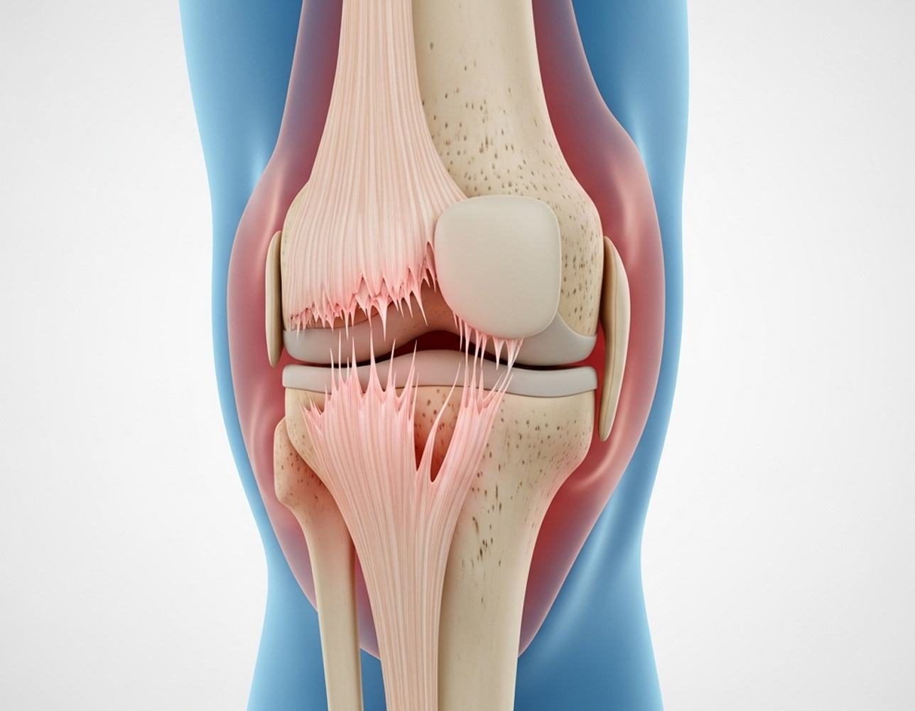
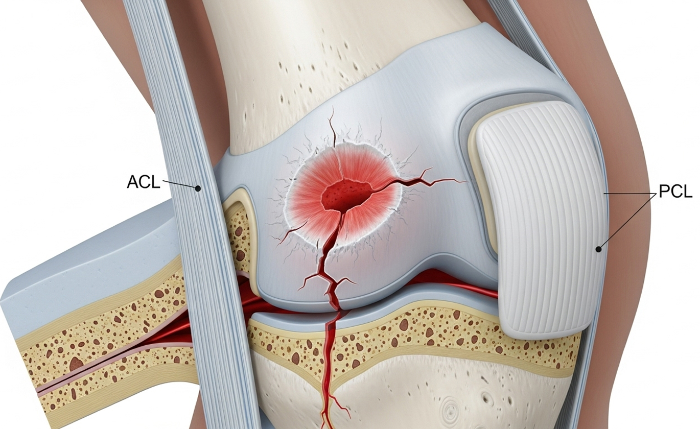
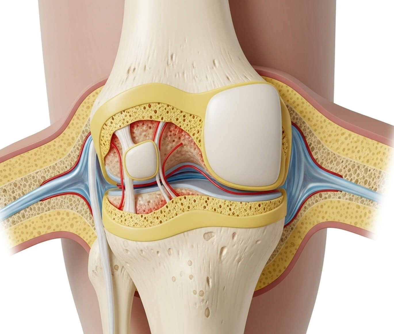
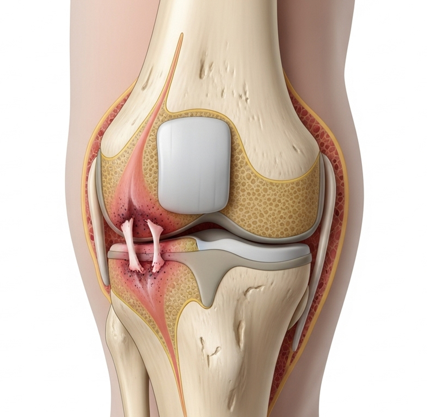
An osteochondral injury involves damage to both the articular cartilage and the underlying bone in a joint, most commonly the knee. Think of articular cartilage as the smooth, white tissue that caps the ends of your bones within a joint, allowing them to glide frictionlessly. Beneath this cartilage lies the subchondral bone, which provides crucial support.

This type of cartilage bone injury knee is more complex than damage limited only to the cartilage surface because it affects this integrated cartilage-bone unit. The term 'osteochondral' itself tells the story: 'osteo' refers to bone, and 'chondral' refers to cartilage.

Understanding Your Knee Cartilage

Articular Cartilage

This smooth, resilient tissue is a specific type called hyaline cartilage. It acts like a shock absorber during activities like walking or running and enables your knee to bend and straighten smoothly. Crucially, it has no direct blood supply ('avascular'). It relies on joint fluid for nourishment, which means it has very limited ability to heal itself if damaged.

Subchondral Bone

This is the layer of bone just beneath the cartilage. It provides structural support. Unlike cartilage, it has a blood supply ('vascular'), giving it a better capacity for healing. However, damage here can compromise the overlying cartilage.

How Common are Osteochondral Injuries?

While not extremely common, osteochondral injuries are significant when they occur, particularly in active people. They are seen more often in younger individuals, typically between 15 and 30 years old, often due to sports trauma. Statistics suggest they can occur alongside 5-10% of ACL injuries and are present in 20-30% of kneecap dislocations. These injuries account for roughly 5-10% of all knee arthroscopies (keyhole surgeries) performed.

Within the knee, these injuries most frequently happen on the end of the thigh bone (femur), specifically:
  • The outer part (lateral femoral condyle), often linked to patella dislocations (50-60% of cases).
  • The inner part (medial femoral condyle) (30-40%).
  • Less commonly, the kneecap (patella) (10-20%) or the top surface of the shin bone (tibial plateau).

Causes and Mechanisms

Osteochondral injuries can happen suddenly due to trauma or develop more gradually.

Traumatic Osteochondral Injuries

These typically result from significant force:

Patella Dislocation (30-40% of cases)

When the kneecap shifts out of place (usually outwards), the inner edge of the kneecap can impact the outer part of the thigh bone. This shearing force can knock off a fragment of cartilage and bone. If the bone piece is large enough, it might be visible on an X-ray.

ACL or Other Ligament Injuries (20-30%)

Severe twisting or pivoting movements that tear ligaments like the ACL can also cause bone bruising and damage the cartilage and underlying bone. These injuries often require an MRI scan for accurate diagnosis.

Direct Knee Trauma

A direct blow to the knee, such as hitting the dashboard in a car accident, a collision in sport, or falling directly onto a bent knee, can cause an osteochondral fracture.

Non-Traumatic or Degenerative Causes

Osteochondritis Dissecans (OCD)

This condition involves the subchondral bone losing its blood supply in a small area, potentially causing it to soften or collapse. This instability affects the overlying cartilage, which can then crack or detach.
  • Juvenile OCD (ages 10-20): Occurs in growing individuals and sometimes heals without surgery.
  • Juvenile OCD (ages 10-20): Occurs in growing individuals and sometimes heals without surgery.

Repetitive Microtrauma

Overuse or repetitive stress, particularly in athletes, can lead to cumulative damage to the cartilage and bone over time.

Types of Osteochondral Injuries

Doctors classify osteochondral injuries based on several factors, including the stability of the fragment and its size. This helps guide osteochondral defect treatment.

By Stability

Stable

The damaged fragment is still firmly attached. These may heal without surgery, especially in young patients with OCD.

Unstable

The fragment is partially detached and may move slightly. It's unlikely to heal without intervention.

Displaced

The fragment has completely broken off and is loose within the knee joint (a 'loose body'). This requires surgery.

By Size

Small (less than 1cm²)

May not cause significant symptoms. Treatment might involve microfracture or drilling.

Medium (1-2cm²)

Usually causes symptoms. Options include microfracture, ACI, or osteochondral autograft (OATS).

Large (greater than 2cm²)

Significantly impacts knee function. Often requires ACI or osteochondral allograft transplantation.
The International Cartilage Regeneration & Joint Preservation Society (ICRS) grades cartilage damage from 0 (normal) to IV (full-thickness loss). An osteochondral injury typically corresponds to Grade IV cartilage damage plus underlying bone involvement.

Symptoms and Diagnosis

Recognising the symptoms is the first step towards getting an accurate diagnosis and appropriate care.

Clinical Symptoms

Symptoms can vary depending on whether the injury is sudden (acute) or has developed over time (chronic).

Acute Symptoms (Sudden Injury)

  • Sudden, sharp pain at the moment of injury.
  • Rapid swelling of the knee, often within hours. If a fragment breaks loose, bleeding into the joint (haemarthrosis) can cause significant swelling.
  • Mechanical symptoms if a piece is loose:
  • Catching or locking sensation during movement.
  • Clicking or popping sounds.
  • Sudden sharp pain with certain movements.
  • Giving-way episodes, where the knee feels unstable or buckles.
  • Difficulty putting weight on the affected leg.

Chronic Symptoms (Gradual Onset or Untreated Injury)

  • Persistent aching pain, often localised to the area of damage.
  • Pain that worsens with activity, especially weight-bearing, climbing stairs, or squatting.
  • Swelling that occurs after activity.
  • Stiffness, particularly in the morning or after periods of rest.
  • Mechanical symptoms (locking, catching) if a fragment becomes loose over time.
  • Gradual loss of function and ability to participate in activities.

Physical Examination

During your consultation, I will perform a thorough examination of your knee. This involves:
  • Assessing for swelling or fluid in the joint (effusion).
  • Checking for tenderness over specific areas of the joint line, which can indicate the defect's location.
  • Evaluating your knee's range of motion and checking for any mechanical blocks or clicks.
  • Performing specific tests, like the Wilson test, which can suggest OCD on the medial femoral condyle.
  • Checking ligament stability if an associated injury like an ACL tear is suspected.

Imaging

Imaging is essential for confirming the diagnosis and planning treatment.

X-Rays

Often the first imaging test. X-rays can show bony fragments that may have broken off or reveal signs of OCD. Specific views (like tunnel or Merchant views) help visualise certain areas of the knee better.

CT Scan

Sometimes used for a more detailed look at the bony anatomy, especially for complex fractures or when planning surgery involving bone grafts. 3D reconstructions can be helpful.

MRI Scan

This is the gold standard for diagnosing osteochondral injuries. MRI provides detailed images of both cartilage and bone, allowing assessment of the lesion's size, exact location, and stability. It also shows any associated injuries, such as ligament tears or meniscal damage. MRI findings are crucial for surgical planning.

Arthroscopy

Keyhole surgery can be used both for diagnosis and treatment. It allows direct visualisation of the cartilage surface and is the most accurate way to assess the stability of a fragment. If needed, treatment can often be performed during the same procedure.

Conservative Treatment

Surgery isn't always necessary for osteochondral injuries. In certain situations, a non-surgical approach might be appropriate.

Indications for Non-Surgical Management

Conservative treatment may be considered for:
  • Stable lesions where the fragment hasn't moved.
  • Juvenile OCD in patients under 15 with open growth plates, as these have a good chance of healing naturally.
  • Small lesions (less than 1cm²) causing minimal symptoms.
  • Injuries found incidentally on imaging that aren't causing symptoms.
  • Older individuals or those with lower activity demands.

Conservative Management Protocol

A typical non-surgical plan includes:

Activity Modification

Reducing or stopping high-impact activities (running, jumping) and switching to low-impact options like swimming or cycling.

Protected Weight-Bearing

Using crutches for a period (perhaps 2-6 weeks) if symptoms are severe, followed by gradual return to full weight-bearing.

Physiotherapy

A tailored program focusing on strengthening the quadriceps muscles, maintaining range of motion, and improving balance (proprioception).

Medications

Simple pain relief like paracetamol, or short-term use of non-steroidal anti-inflammatory drugs (NSAIDs) to manage pain and swelling.

Monitoring

Regular follow-up appointments with repeat X-rays or MRI scans (usually every 3-6 months) to track healing or detect any worsening.

Conservative Treatment Outcomes

Success rates depend largely on the type of injury and the patient's age:

Juvenile OCD (under 15

Healing occurs in 50-70% of cases with conservative management, especially if growth plates are still open. Treatment typically lasts 6-12 months. Surgery may be needed if there's no sign of healing by then.

Adult Stable OCD or Small Lesions

While 30-50% of adults may experience symptom improvement with activity changes, actual healing of the defect is rare due to cartilage's limited blood supply. Many may eventually require surgery.

Displaced or Unstable Lesion

Conservative treatment is generally ineffective for these injuries, and surgery is usually required.

Surgical Treatment Options

When conservative treatment isn't appropriate or hasn't succeeded, or if the injury is unstable or displaced, surgery offers various options for osteochondral defect treatment and knee cartilage repair. My approach is always evidence-based, considering the specifics of your injury and your goals.

Arthroscopic Assessment and Fragment Removal

For loose bodies or unstable fragments causing mechanical symptoms like locking or catching, keyhole surgery (arthroscopy) can be used to remove the problematic piece. While this often improves symptoms significantly, the underlying defect in the cartilage and bone remains. This is generally considered a palliative measure to reduce symptoms rather than a restorative repair.

Osteochondral Fragment Fixation

If a large fragment (usually >1cm²) breaks off acutely (within about 6 weeks of injury) and is otherwise healthy, it may be possible to fix it back in place. This is done arthroscopically or through a small incision, using specialised screws or bioabsorbable implants. If the fragment heals successfully (around 60-80% healing rate), this preserves your natural cartilage surface.

Microfracture / Drilling

This is often a first-line treatment for small to medium-sized defects (less than 2cm²). Performed arthroscopically, small holes are created in the exposed subchondral bone at the base of the defect. This stimulates bleeding and the formation of a clot rich in marrow cells, which eventually forms a type of scar cartilage called fibrocartilage.

Microfracture is a single-stage procedure with relatively low cost and complication rates. Good short-term results (2-5 years) are seen in 60-80% of patients. However, fibrocartilage is less durable than the original hyaline cartilage and tends to wear down over 5-10 years.

Osteochondral Autograft Transfer (OATS / Mosaicplasty)

Suitable for medium-sized defects (1-2.5cm²) in younger, active patients. This procedure involves harvesting small cylindrical plugs of healthy cartilage and bone from a non-weight-bearing area of your own knee (autograft). These plugs are then transferred and press-fitted into the defect site, creating a mosaic-like surface.

The main advantage of OATS is that it restores the defect with your own hyaline cartilage, offering good long-term durability (often 10 years or more). Success rates are generally high, around 75-90% good outcomes at 5-10 years. Limitations include the amount of graft available from the donor site and potential pain or issues at the harvest site.

Autologous Chondrocyte Implantation (ACI)

ACI is typically reserved for larger defects (greater than 2cm²) or situations where other surgeries have failed, usually in younger patients (under 40). It's a two-stage procedure:

  1. Stage 1: A small biopsy of healthy cartilage is taken from your knee arthroscopically. These cartilage cells (chondrocytes) are grown and multiplied in a laboratory over 3-6 weeks.
  2. Stage 2: A second, open surgery is performed. The cultured cells are implanted into the defect, often held in place under a special membrane or scaffold, where they regenerate hyaline-like cartilage.

ACI can treat large defects (up to 10cm²) and achieves good long-term outcomes (70-85% at 10+ years). However, it involves two surgeries, is expensive, has limited public funding in Australia, and requires prolonged rehabilitation (12-18 months).

Osteochondral Allograft Transplantation

Used for very large defects (often >2.5-3cm²), particularly those with significant underlying bone loss, or as a revision option after other failed procedures. This involves transplanting a size-matched graft of cartilage and bone harvested from a deceased donor (allograft). It's a single-stage procedure.

Advantages include the ability to treat very large defects without donor site issues. Disadvantages include limited graft availability, the potential for disease transmission (though rigorously screened), and a risk of graft rejection or failure over time (10-25% at 5-10 years). Graft survival rates are around 65-85% at 10 years.

Recovery and Outcomes

Rehabilitation after surgery is crucial for a successful outcome and varies depending on the procedure performed.

Rehabilitation Timeline

Each procedure requires a specific, structured physiotherapy programme. General timelines are:

Microfracture

Typically 6 weeks of non-weight-bearing or protected weight-bearing on crutches, followed by gradual progression. Return to sport may take 6-9 months.

OATS

Weight-bearing progression usually starts earlier, often within 2-6 weeks. Return to sport is typically around 6-9 months.

ACI

Requires a longer period of protected weight-bearing, often 6 weeks, sometimes using a continuous passive motion (CPM) machine to gently move the knee.

Allograft

Similar protected weight-bearing phase to ACI, with return to sport generally guided by graft incorporation and functional recovery, often 9-12 months or longer.

Return-to-Sport Expectations

Returning to sport after knee cartilage repair depends on the procedure, defect size, sport type, and individual recovery.

  • Return rates vary: Microfracture (60-75%), OATS (75-85%), ACI (70-80%), Allograft (65-80%).
  • While many patients return to recreational sports, achieving the pre-injury level of performance, especially in high-impact or competitive sports, can be challenging.
  • Factors influencing return include defect size, surgical technique, rehabilitation adherence, age, and type of sport. Lower-impact sports generally have higher return rates.
  • I discuss realistic expectations for returning to sport with each patient during consultation, based on their specific injury and goals.

Dr Allom's Approach to Osteochondral Injuries

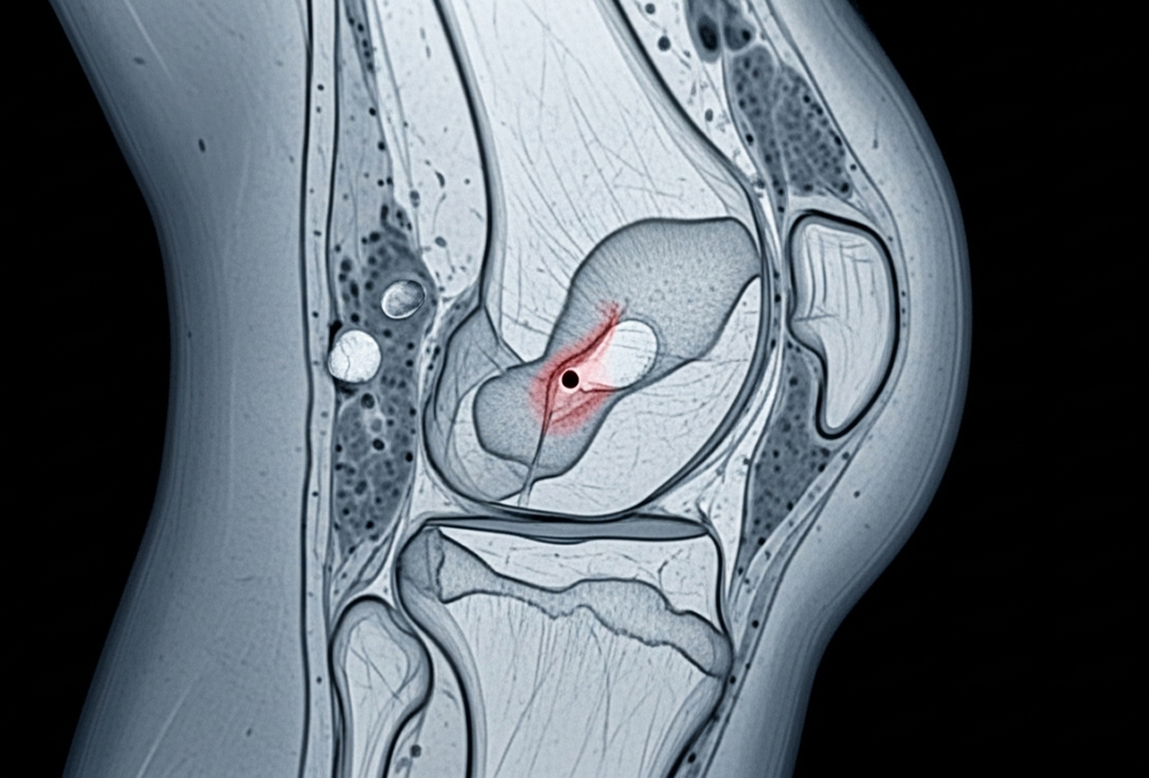
Treating osteochondral injuries requires careful evaluation and a tailored approach. As a fellowship-trained knee subspecialist, I provide comprehensive assessment through clinical examination and detailed review of your imaging (X-ray and MRI). When necessary, diagnostic arthroscopy allows direct visualisation.

My treatment recommendations are always based on the best available evidence, considering the specifics of your osteochondral injury knee, your age, activity level, and long-term goals. Depending on your situation, this may involve a trial of conservative management, arthroscopic surgery for fragment fixation or removal, microfracture, or OATS (mosaicplasty). For very large defects requiring ACI or allograft, I can provide assessment and referral to specialised centres when indicated.

Successful recovery hinges on precise, procedure-specific rehabilitation. I work closely with experienced physiotherapists to ensure you follow the optimal protocol for your surgery, guiding your gradual return to activity. My dual fellowship training (FRCS from the UK and FRACS from Australia) and dedicated focus on knee surgery provide the subspecialist expertise needed to manage these complex injuries effectively.

Frequently Asked Questions

In adults, it's unlikely for a significant osteochondral injury to heal completely without intervention. Cartilage has very limited healing capacity due to its lack of blood supply.
  • Juvenile OCD (under 15): Has the best chance, with 50-70% potentially healing with rest and activity modification.
  • Adult Stable Lesions: Symptom improvement occurs in less than 30% without surgery, but true healing is rare.
  • Unstable or Displaced Fragments: These do not heal without surgical fixation or removal.
A trial of conservative management is reasonable for stable juvenile OCD, but most adult osteochondral injuries eventually require surgery for optimal long-term outcomes.

There isn't a single "best" surgery; the ideal osteochondral defect treatment is highly individualised. Factors influencing the decision include:

  • Defect Size: Smaller defects (<1cm²) might suit microfracture; medium (1-2.5cm²) often suit OATS; larger (>2.5cm²) typically require ACI or allograft.
  • Age: Younger, active patients (<40) are generally better candidates for restorative procedures like OATS or ACI. Older or lower-demand individuals might opt for microfracture or fragment removal.
  • Activity Level: High-demand athletes often benefit more from OATS or ACI which restore more durable cartilage. Microfracture may suffice for lower-demand lifestyles.
  • Previous Surgeries: If a microfracture has failed, ACI or OATS might be considered. Failed OATS might lead to ACI or allograft.
I will recommend the most appropriate option based on a thorough assessment of your imaging, symptoms, and personal goals.

Durability varies significantly by procedure:
  • Microfracture: Good results often last 2-5 years, but the fibrocartilage can deteriorate over 5-10 years.
  • OATS: Generally durable for 10-15 years or more, as it uses your own hyaline cartilage.
  • ACI: Can provide durable results for 10-15+ years when used for appropriate large defects.
  • Osteochondral Allograft: Durability is variable, typically 5-15 years, with graft failure risk increasing over time.
Factors like surgical technique, rehabilitation compliance, activity modification, weight management, and defect location also impact long-term success.

It's possible for some, but not guaranteed, and depends on many factors. Favourable factors include:
  • Smaller defect size.
  • Younger age (under 30).
  • Excellent rehabilitation.
  • Type of sport (lower impact sports have better return rates).
Challenges include the repaired cartilage potentially not withstanding repetitive high impact, risk of re-injury, and often a reduced performance level compared to before injury. Many patients successfully return to recreational sport, but competitive high-impact sport remains challenging.

Yes, unfortunately, having an osteochondral injury increases your risk of developing osteoarthritis in that knee later in life, even with successful treatment. The goal of treatment is to restore the joint surface as well as possible to minimise this risk. Strategies to reduce arthritis progression include timely treatment, maintaining a healthy weight, modifying high-impact activities, and keeping your thigh muscles (quadriceps) strong.

Symptoms typically include sudden episodes of the knee locking or catching, sharp intermittent pain, or a sensation of something moving around inside the joint. Diagnosis is confirmed with imaging (X-ray or MRI) or during arthroscopy. Arthroscopic removal of the loose body usually resolves these mechanical symptoms effectively.

Currently, there are no proven non-surgical treatments that reliably regenerate damaged articular cartilage. Supplements like glucosamine have limited evidence, and the effectiveness of injections like Platelet-Rich Plasma (PRP) or stem cells for cartilage regeneration remains unproven or is still under investigation. Surgical procedures like ACI, OATS, and microfracture remain the only established methods for stimulating cartilage repair.

Next Steps

If you have symptoms of an osteochondral injury or have been diagnosed with cartilage and bone damage in your knee, I encourage you to schedule a consultation. Together, we can discuss your situation and determine the best treatment path for you.

What to Bring: Please bring your referral letter, all relevant imaging studies (X-rays, CT scans, MRI if performed), and your list of current medications to your consultation.

Convenient Locations in Two Regions

I consult and operate from five locations, providing local access to subspecialist expertise without the need for long-distance travel.

South West Sydney Locations

Phone Number

04 3818 3832

Gledswood Hills

The George Centre, Suite 12, 1A The Hermitage Way

Campbelltown

Centric Park, Level 3, 4 Hyde Parade

Liverpool

Sydney South West Private Hospital, Suite 3.02, 24-40 Bigge Street

Mid North Coast Locations

Phone Number

02 6551 0722

Taree

Mayo Private Hospital Specialist Suites, 2 Potoroo Drive

Forster

Dolphin Suites Consulting Rooms, 33 Breckenridge Street

Book Your Consultation

©2026 Dr Richard Allom. All rights reserved.

Oxford Knee Score Questionnaire

Please answer the following 12 questions about your knee, considering your symptoms over the past 4 weeks. Select one answer for each question.